Showing posts with label Surgery. Show all posts

6 Jan 2013

Blood circulations

Blood circulations

There are three types of circulation :

  1. Pulmonary circulation :

    - Venous blood passes from the right ventricle through the pulmonary trunk to the lungs to be oxygenated.
    -Oxygenated blood returns from the lungs through 4 pulmonary veins to the left atrium.


  2. Systemic circulation :

    - Oxygenated blood passes from the left ventricle through the aorta and its branches to supply the different tissues of the body.
    - Venous blood returns from the tissues of the body along small veins which unites to form superior vena cava, inferior vena cava and coronary sinus which open into the right atrium.


  3. Portal circulation :

    - The portal vein drains the venous blood from stomach, intestines, spleen and pancreas and passes into the liver.
    - This blood carries the products of digestion to the liver.
    - The portal vein divides repeatedly inside the liver to end in the liver sinusoids.
    - The liver sinusoids lead to hepatic vein which join inferior vena cava.
    - Thereafter, blood in the inferior vena cava enters the right atrium.


5 Jan 2013

Connections between arteries and veins

Connections between arteries and veins


The arteries maybe connected to veins by one of the following methods :
  1. Capillaries > which connect the arterioles with the venules.
  2. Arterio-venous anastomosis > these are direct connections between small arteriols and small venules
  3. Sinusoids > these are irregular wide spaces connecting the arteries and veins.

Capillaries


Arterio-venous anastomosis


 Sinusoids


Types of muscles

Types of muscles

Skeletal muscles
Cardiac muscle
smooth muscles

Attached to skeleton
In myocardium
In internal viscera and blood vessels
Distribution
Voluntary
Involuntary
Involuntary
Contraction
Present
Present
Absent
Striations
Somatic
Autonomic
Autonomic
Nerve supply
Multinuclear
Branched and fused together with single nucleus
Spindle-shaped with one nucleus
Muscle cell


Image

Synovial joints

Synovial joints

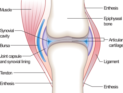
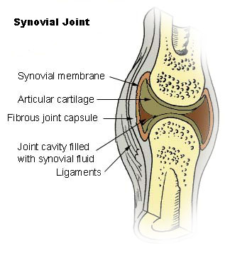
* Structure :

- The joint is enveloped by fibrous capsule.
- The capsule is thickened in some places forming ligaments.
- The capsule is lined with synovial membrane.
- The articular surface of the bone is covered by articular cartilage.
- The joint cavity contains synovial fluid.
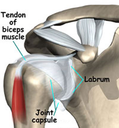
- The joint cavity may contain other additional intracapsular structures like 
   inter-articular disc, labrum, meniscus or tendons of muscles.

* Classification :

( According to the number of axes around which movements occur )

  1. Uni-axial joints ( one axis ) :
    1. Hinge joints : transverse axis > flexion and extension > elbow joint
    2. Pivot joints : vertical axis > rotation > radio-ulnar joint
  2. Bi-axial joints ( two axes ) :
    1. Ellipsoid joints : flexion, extension, adduction and abduction > wrist joint 
    2. Saddle joints : flexion, extension, adduction, abduction and slight rotation > carpo-metacarpal joint of the thumb.
  3. Multi-axial joints ( more than two axes ) :All types of movement are allowed > shoulder and hip joints.
  4. Other types :
    1. Plane joints : the articular surface are flat > just gliding > intercarpal joints.
    2. Compound joints : more than one type in the same joint > tempro-mandibular joint.


* Illustrations :

Structure of synovial joints :


Inter-articular disc


Labrum


Meniscus

Hinge joint

Pivot joint


Ellipsoid joint


Saddle joint


Multi-axial joint


Plane joint

Compound joint